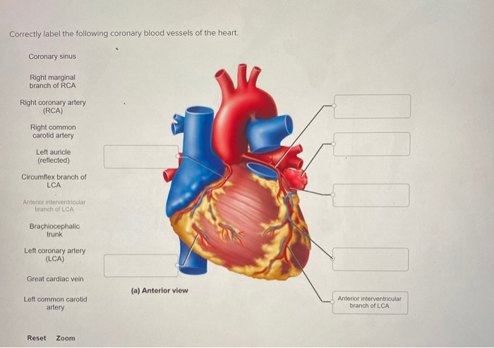
Correctly Label the Following Coronary Blood Vessels of the Heart.
Carries deoxygenated blood from the body to the heart. Correctly label the pathway for the cardiac conduction system.
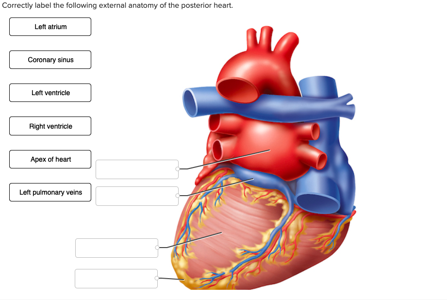
Solved Correctly Label The Following External Anatomy Of The Chegg Com
Carries oxygenated blood from the lungs to the heart.

. The left descending artery supplies blood to the front left side of the heart and circumflex artery supplies the outside and the back of the heart with blood. This system is a network of blood vessels such as arteries veins and capillaries that carries blood to and from all areas of your body. Correctly label the following coronary blood vessels of the heart.
Right coronary artery RCA - supplies blood to the right atrium right ventricle bottom portion of the left ventricle and back of the septum. Two major coronary arteries branch off from the aorta near the point where the aorta and the left ventricle meet. The largest arteries have special elastic fibres in their walls.
Blood vessels leading into and out of the heart. The aorta is the largest artery in the body. They can vary in size.
Pathway of blood through the heart 1. Structure of the heart showing top 8 worksheets in the category structure of the heart. Recall that blood returning from the systemic circuit enters the right atrium Figure 2052 via the superior and inferior venae cavae and the coronary sinus which drains the blood supply of the heart muscleThese vessels will be described more fully later in this section.
Because the rest of the body and most especially the brain needs a steady supply of oxygenated blood that is free of all but the slightest interruptions the. There are five main types of blood vessels. Right and left pulmonary arteries 8.
The heart is made of cardiac muscle composed of cells called myocytes. The heart is an organ about the size of your fist that pumps blood through your body. Coronary arteries supply oxygenated blood to the heart muscle.
Arteries play a major role in nourishing organs with blood and nutrients. UNIT 12 QUIZ Name continued Date Section 8 Label the following arteries in Figures 1234 and 1235. The left ventricle then pumps blood through the aortic valve and into the aorta.
It carries oxygenated. Structure Of The Heart Worksheet. Categorize the following changes with regard to how they will impact cardiac output.
Pulmonary veins -bring oxygen-rich blood back to the heart from the lungs. It is made up of multiple layers of tissue. Here is a Heart labeling quiz for you.
Flaps that prevent backflow of blood. Superior and inferior vena cavae and the coronary sinus 2. From the right ventricle it goes through the pulmonary semilunar valves to the pulmonary trunk 4.
The right and the left. Left Anterior Descending artery LAD The left coronary arteries supply. Blood enters the right atrium from the superior and inferior venae cavae and the coronary sinus.
Figure 1234 OBrachial artery Femoral artery OLeft common carotid artery O Posterior tibial artery Radial artery ORenal artery ORight subclavian artery O Ulnar artery O Vertebral artery FIGURE 1234 Major arteries of the body Figure 1235 O Celiac trunk O. Carries oxygenated blood from the heart around the body. Correctly label the following coronary blood vessels of the heart.
The presence of elastin in the large blood vessels enables these vessels to increase in size and alter their diameter. Receives oxygenated blood from the lungs. Correctly label the great vessels that enter and exit the heart.
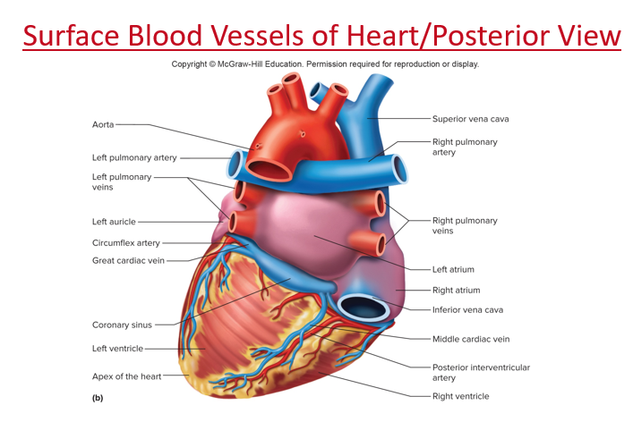
Correctly label the following coronary blood vessels of the heart. These arteries and their branches supply all parts of the heart muscle with blood. Coronary circulation is the circulation of blood in the blood vessels that supply the heart muscle.
The aorta is the only structure in the list that carries oxygenated blood. However the pulmonary vein carries oxygenated blood to the heart. The arteries carry the blood rich in oxygen from the heart to different parts of the body.
Arteries arterioles capillaries venules and veins. Together with the left anterior descending. Coronary sinus Right marginal branch of RCA Right coronary artery RCA Right common carotid artery Left auricle reflected Circumflex branch of LCA Anterior interventricular branch of LCA Brachiocephalic trunk Left coronary artery LCA Great cardiac vein a Anterior view Left common.
Tricuspid valve also called the rt. The right coronary artery supplies the right side the right ventricle right atrium sinoatrial node SA. Right coronary artery RCA.
Region of the heart that pumps oxygenated blood to the body. Your heart is at the center of your circulatory system. Categorize the following in terms of the muscle type to.
The pulmonary artery being an exception carries deoxygenated blood to the lungs for purification. Cardiac veins then drain away the blood after it has been deoxygenated. Atrioventricular valve or mitral vale 10.
Drag each label to the location of each structure described. The veins carry impure blood from different parts of the body to the heart for oxygenation. To accommodate this stress they have an abundance of elastic tissue and less smooth muscle.
Take the following quiz to know how much you know about your heart. Pulmonary semilunar valve 6. Left Main Coronary Artery also called the left main trunk The left main coronary artery branches into.
The right coronary artery supplies blood to the right ventricle the right atrium and the SA sinoatrial and AV atrioventricular nodes which regulate the heart rhythm. There are two main coronary arteries. This blood carries little oxygen as it is returning from the body where.
There are four main blood vessels that take blood into and out of the heart. Arteries are always under high pressure. Arteries carry blood away from the heart to other organs.
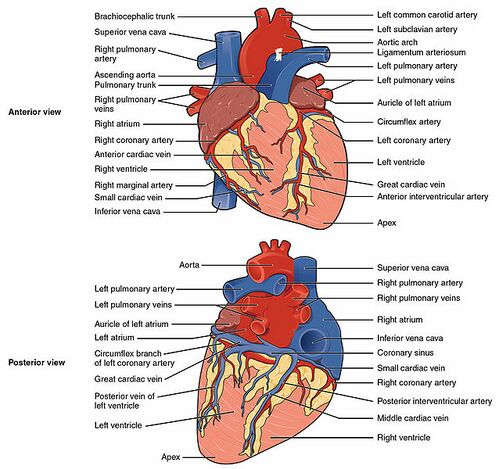
The major blood vessels that are connected to the heart include the aorta the superior vena cava the inferior vena cava the pulmonary artery which takes oxygen-poor blood from the heart to the. From right atrium it goes through the tricuspid valve to the right ventricle. Network of blood vessels that branch off the aorta to supply the heart muscle with oxygen-rich blood.
Carries deoxygenated blood to the lungs. The aorta is the artery that feeds the rest of the body through a system of blood vessels. Left anterior descending artery LAD - supplies blood to the front and bottom of the left ventricle and the front of the septum.
Blood returns to the heart from the body via two large blood vessels called the superior vena cava and the inferior vena cava. The more healthy your heart is the longer the chances you have of surviving so you better take care of it. The human heart is a vital organ for every human.
Name the major vessels that leave the heart and identify where they lead to. The right coronary artery divides into smaller branches including the right posterior descending artery and the acute marginal artery. Blood vessels are found throughout the body.
Bicuspid valve also called the lt.

A P Anatomy Physiology The Unity Of Form And Function Chapter 19 The Heart Megakit Diagram Quizlet

Coronary Arteries Radiology Reference Article Radiopaedia Org

Laser System In The Medical Device Industry

A P Anatomy Physiology The Unity Of Form And Function Chapter 19 The Heart Megakit Diagram Quizlet

Chapter 20 Cardiovascular System Flashcards Quizlet

Ahcdw15notes11 Pdf 11 Award 1 00 Point Problems Adjust Credit For All Course Hero

Chapter 20 Cardiovascular System Flashcards Quizlet
Coronary Vessels Quiz By Thebrend88
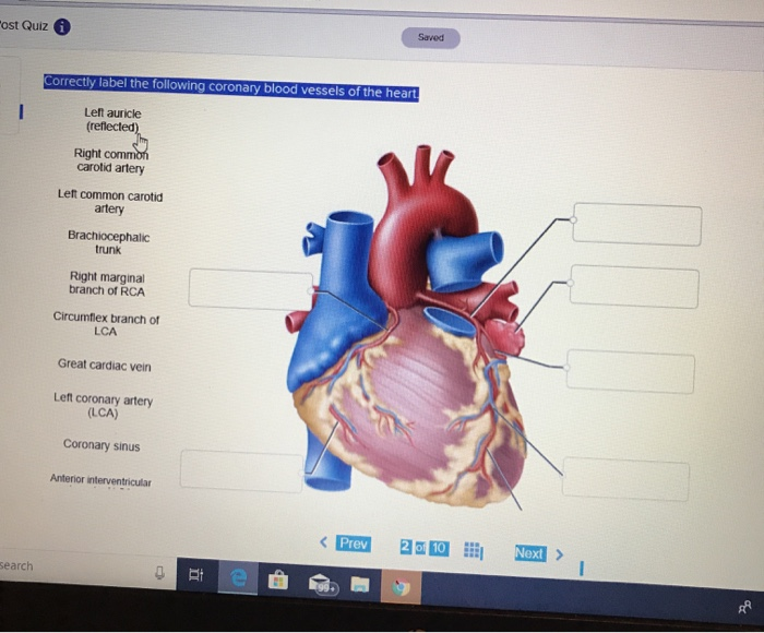
Solved Saved Ost Quiz I Correctly Label The Following Chegg Com

Ahcdw15notes21 Pdf 21 Award 1 00 Point Problems Adjust Credit For All Students Correctly Label The Following Coronary Blood Vessels Of The Course Hero
Solved Correctly Label The Following Internal Anatomy Of The Chegg Com
Solved Correctly Label The Following External Anatomy Of The Posterior Heart Course Hero

Heart Anatomy Chambers Valves And Vessels Cardiac Anatomy Heart Arteries Heart Diagram
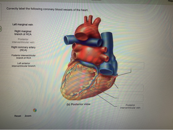
Solved Help Correctly Label The Following Coronary Blood Chegg Com
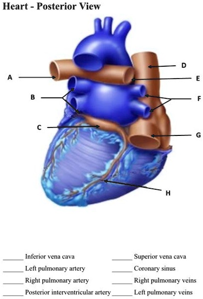
Solved Heart Posterior View Inferior Vena Cava Superior Vena Cava Coronary Sinus Right Pulmonary Veins Left Pulmonary Veins Left Pulmonary Artery Right Pulmonary Artery Posterior Interventricular Artery

Ahcdw15notes12 Pdf 12 Award 1 00 Point Problems Adjust Credit For All Students Correctly Label The Following External Anatomy Of The Posterior Course Hero
Anatomy Of The Human Heart Physiopedia
Solved Correctly Label The Following Coronary Blood Vessels Chegg Com
Ch 13 Cardiovascular System Biology Quizizz


Comments
Post a Comment